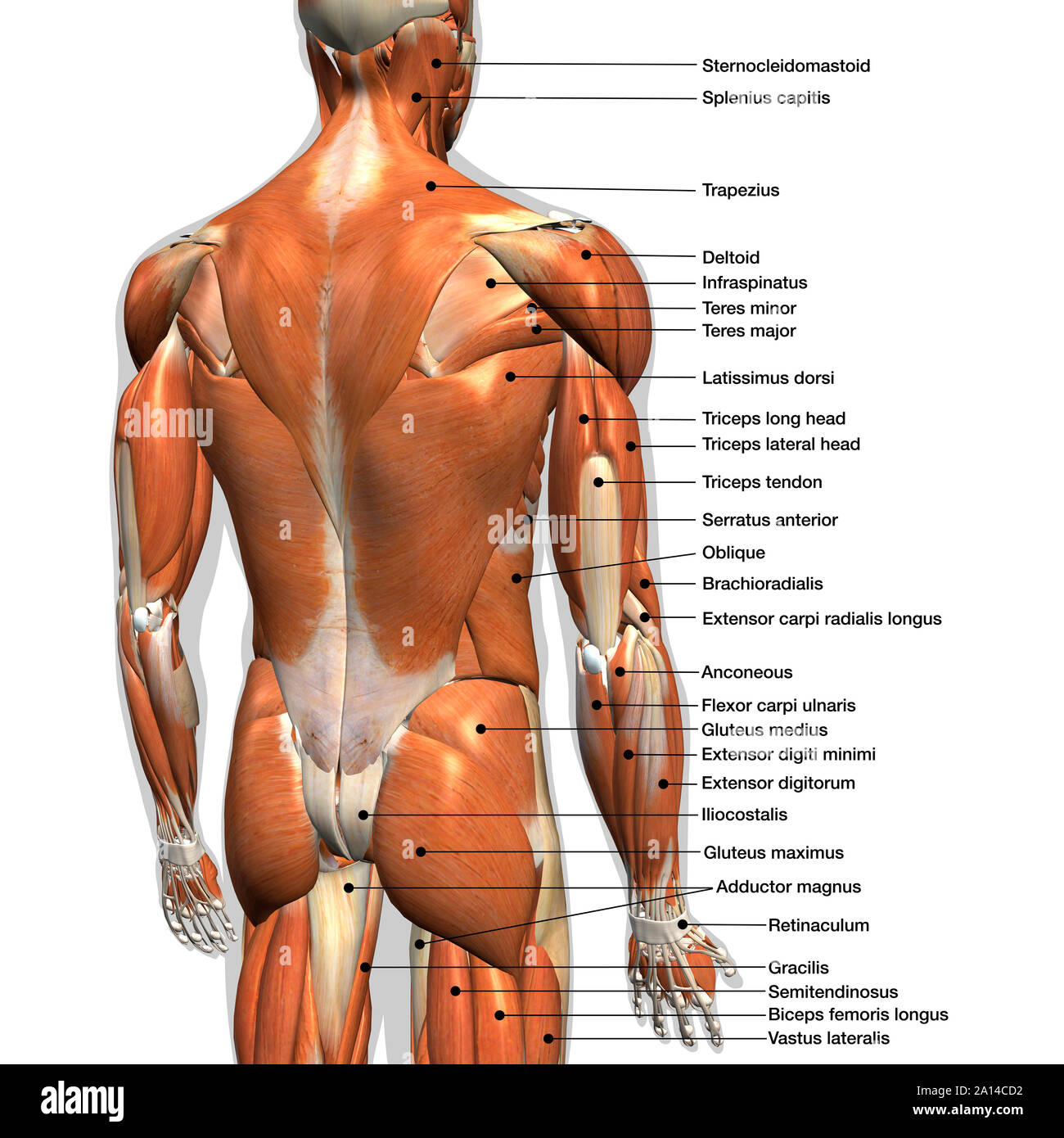
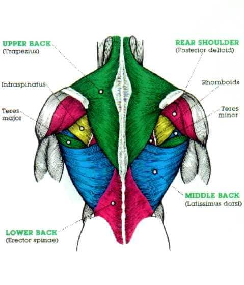
Back Muscles Anatomy : 1) make midline incision along spines of vertebrae 2) extend from. 1) make midline incision along spines of vertebrae 2) extend from If we want to locate the back muscles in the body, we can say that it starts from the top of the neck and ends at the bottom of the pelvis. Balance the weight of your head on top of your spine evenly distribute weights from your upper body into the lower extremities Anatomy of back muscles your back consists of three distinct layers of muscles, namely the superficial layer, the intermediate layer, and the deep layer. The trapezius and latissimus dorsi muscles connect the upper limb to the vertebral column.
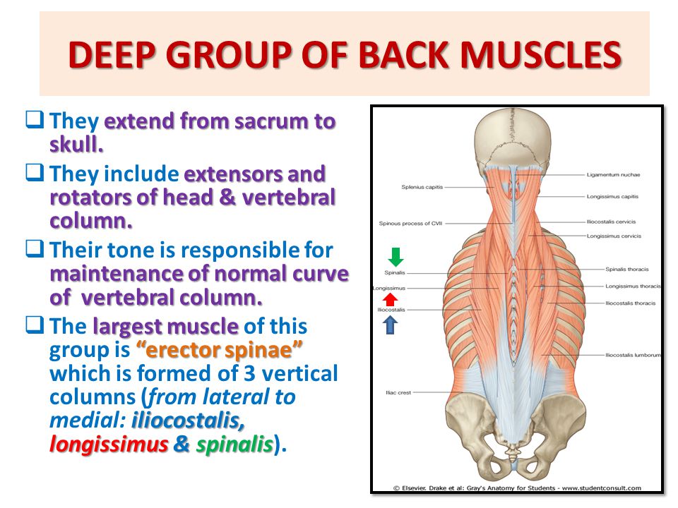
Superiorly and the latissimus dorsi m. All about the back muscles the back anatomy includes the latissimus dorsi, trapezius, erector spinae, rhomboid, and the teres major. The back muscles are anatomically layered into superficial (extrinsic) and deep (intrinsic) muscles. The back muscles are divided into two large groups: Its floor is the posterior thoracic wall
Labeled Anatomy Chart Of Male Back Muscles On White Background Stock Photo Alamy from c8.alamy.com The superficial back muscles are situated underneath the skin and superficial fascia. They provide movements of the spine , stability to the trunk, as well as the coordination between the movements of the limbs and trunk. Superiorly and the latissimus dorsi m. Topographic anatomy of the back; They start at the top of the neck and go down to the tailbone. Anatomy chart courtesy of fcit the latissimus dorsi muscles (also known as the lats) are the largest muscles of the back. Muscle or ligament strains can occur from repeated use of the muscles, or from improperly or awkwardly lifting heavy objects. All about the back muscles the back anatomy includes the latissimus dorsi, trapezius, erector spinae, rhomboid, and the teres major.
Browse 3,579 back muscle anatomy stock photos and images available, or search for pelvic bone or lymphatic system to find more great stock photos and pictures.
Muscle or ligament strains can occur from repeated use of the muscles, or from improperly or awkwardly lifting heavy objects. Here is everything you need to know to build an unbelievable back. Back pain is common and might be caused by a problem with a muscle. These muscles work together to move the scapula anteriorly and laterally during pushing, throwing, or punching motions. These sections are cervical (neck), thoracic (upper and middle back), lumbar (lower back), and sacrum (tailbone). On this page, you'll learn about each of these muscles, their locations and functional anatomy. Anatomy chart courtesy of fcit the latissimus dorsi muscles (also known as the lats) are the largest muscles of the back. All these muscles are therefore associated with movements of the upper limb. Back muscles chart, find out more about back muscles chart. Your lower back (lumbar spine) is the anatomic region between your lowest rib and the upper part of the buttock. Since the all the back muscles originate in embryo (fetus) form by locations other than the back, muscles in the superficial, as well as, intermediate groups, are extrinsic muscles. Artery) p.134 accessory nerve p. These structures work together to support the body, enable a range of movements, and send messages from the brain to the.
Anatomy chart courtesy of fcit the latissimus dorsi muscles (also known as the lats) are the largest muscles of the back. It is bounded by the trapezius m. The muscles, bones, ligaments, and tendons in the back can all be injured and cause back pain. Muscular anatomy of the back. If we want to locate the back muscles in the body, we can say that it starts from the top of the neck and ends at the bottom of the pelvis.
Muscles Of Back Prof Ahmed Fathalla Ibrahim Professor Of Anatomy Ppt Video Online Download from slideplayer.com It is bounded by the trapezius m. Since the all the back muscles originate in embryo (fetus) form by locations other than the back, muscles in the superficial, as well as, intermediate groups, are extrinsic muscles. Three types of back muscles that help the spine function are extensors, flexors and obliques. All these muscles are therefore associated with movements of the upper limb. On this page, you'll learn about each of these muscles, their locations and functional anatomy. These muscles give height and breadth to back development. Browse 3,565 back anatomy muscles stock photos and images available, or start a new search to explore more stock photos and images. Topographic anatomy of the back;
Balance the weight of your head on top of your spine evenly distribute weights from your upper body into the lower extremities
Canine muscle anatomy 12 photos of the canine muscle anatomy canine muscle anatomy, canine muscle anatomy diagram, canine muscle anatomy model, canine shoulder muscle anatomy, dog muscle. Here is everything you need to know to build an unbelievable back. They start at the top of the neck and go down to the tailbone. Balance the weight of your head on top of your spine evenly distribute weights from your upper body into the lower extremities Human anatomy for muscle, reproductive, and skeleton. 1) make midline incision along spines of vertebrae 2) extend from It is bounded by the trapezius m. In fact, the back contains a group of muscles, not one muscle. Browse 3,579 back muscle anatomy stock photos and images available, or search for pelvic bone or lymphatic system to find more great stock photos and pictures. Superficial back muscles, intermediate back muscles and intrinsic back muscles.the intrinsic muscles are named as such because their embryological development begins in the back, oppose to the superficial and intermediate back muscles which develop elsewhere and are therefore classed as extrinsic muscles. The superficial back muscles are situated underneath the skin and superficial fascia. The back consists of the spine, spinal cord, muscles, ligaments, and nerves. If we want to locate the back muscles in the body, we can say that it starts from the top of the neck and ends at the bottom of the pelvis.
It is bounded by the trapezius m. These layers of back muscles help to mobilize and stabilize your trunk during your day to day activities. In fact, the back contains a group of muscles, not one muscle. These sections are cervical (neck), thoracic (upper and middle back), lumbar (lower back), and sacrum (tailbone). Human musculature bodybuilding infographic muscular system vector human anatomy back muscle anatomy bicep male muscular anatomy human body anatomy female female anatomy muscle hamstrings muscle.
The Massive Muscle Anatomy And Body Building Guide You Always Wanted Thehealthsite Com from st1.thehealthsite.com Anterior rami of spinal nerve innervate them. The extensor muscles are attached to back of the spine and enable standing and lifting objects. These structures work together to support the body, enable a range of movements, and send messages from the brain to the. The surface muscles of the upper back include the trapezius muscles (traps) and posterior deltoids. Superficial back muscles, intermediate back muscles and intrinsic back muscles.the intrinsic muscles are named as such because their embryological development begins in the back, oppose to the superficial and intermediate back muscles which develop elsewhere and are therefore classed as extrinsic muscles. It is bounded by the trapezius m. Its floor is the posterior thoracic wall These muscles give height and breadth to back development.
The human spine is composed of 4 sections of vertebrae.
The deep muscles develop in the back called intrinsic muscles. These muscles give height and breadth to back development. Here is everything you need to know to build an unbelievable back. The back muscles are anatomically layered into superficial (extrinsic) and deep (intrinsic) muscles. Canine muscle anatomy 12 photos of the canine muscle anatomy canine muscle anatomy, canine muscle anatomy diagram, canine muscle anatomy model, canine shoulder muscle anatomy, dog muscle. Both the deltoid and the trapezius are firmly attached to the spine of the scapula. In fact, the back contains a group of muscles, not one muscle. If we want to locate the back muscles in the body, we can say that it starts from the top of the neck and ends at the bottom of the pelvis. Browse 3,579 back muscle anatomy stock photos and images available, or search for pelvic bone or lymphatic system to find more great stock photos and pictures. Back pain is one of the most common kinds of pain for adults, and muscle strains are the most common type of back pain. These structures work together to support the body, enable a range of movements, and send messages from the brain to the. These sections are cervical (neck), thoracic (upper and middle back), lumbar (lower back), and sacrum (tailbone). Since the all the back muscles originate in embryo (fetus) form by locations other than the back, muscles in the superficial, as well as, intermediate groups, are extrinsic muscles.


0 Comments